Differences between a Panniculectomy and a Tummy Tuck

Karim Sarhane
Dr. Karim Sarhane

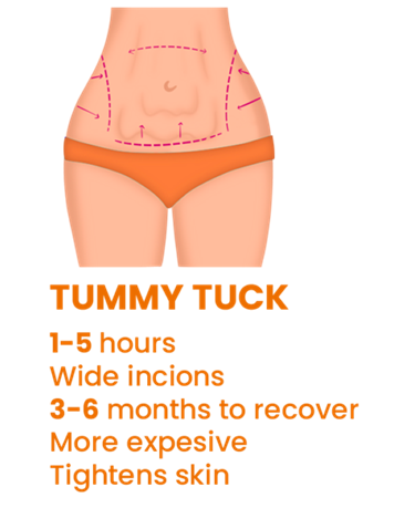
Weight loss helps address health issues like the risk of congenital conditions and can boost self-esteem due to body contouring. The loss, however, can result in loose and sagging skin that does not respond to exercise, and may require removal through surgery to enhance the contouring further. Two primary surgical options exist – panniculectomy and tummy tuck. The choice between the two may be confusing. Though the end goal remains the same, there are differences in the process, candidates, cost, and risks. A panniculectomy involves removing the panniculus, the excess loose skin that forms a flap below the waistline and over the thighs. The panniculus does not emanate from pregnancy, excess weight, or regular weight loss. It emanates from losing excess body weight, typically over 100 pounds, mostly from extensive weight loss procedures like gastric bypass. The extent of a panniculectomy, considered a medical procedure, depends on how much skin is removed. A tummy tuck, on the other hand, is considered a cosmetic procedure that involves removing excess stomach skin and tightening the abdominal muscles. Sometimes surgeons add an abdominal mesh to reinforce muscle repair. This woven mesh surgically temporarily or permanently supports organs after surgery or damaged tissue during healing. The second difference between a panniculectomy and a tummy tuck is eligibility. The former is best for patients with life-impacting sagging skin, which follows heavy weight loss, especially gastric bypass. Other candidates include individuals experiencing hygiene issues under the skin such as ulcers and infections, especially below the pubic region. The excess skin flap can also affect the individual’s mobility. On the other hand, a tummy tuck addresses sagging skin that does not affect the individual’s health and is mainly for cosmetic purposes, especially contouring the body for a more hourglass shape. Other good fits include individuals with sagging skin following a pregnancy who cannot get rid of the excess skin despite exercise and diet changes. In conjunction with liposuction, a tummy tuck can help resolve body-related self-esteem issues and those seeking to get rid of more skin after a panniculectomy. The third difference is the recovery time after each procedure. Recovery from any mild to major surgery involves surgical drains, skin glue, steri-strips, and the dressing. The surgeon provides instructions on how to care for each over the recovery period. A panniculectomy typically takes eight weeks to heal. However, the time may vary depending on the surgery’s extent and the patient’s attendance to the wound after the surgery, especially daily tasks and nutrition. Even weeks after the healing has taken place, however, the patient should refrain from strenuous activities like heavy lifting. Tummy tucks take less time to recover from due to less surgery complexity – usually four to six weeks. With proper care, especially hygiene of the surgical area, the patient should resume regular activity, including strenuous tasks, in the first few weeks following the procedure. Due to the extent of the surgery, panniculectomies are more costly than tummy tucks. A procedure typically costs between $8,000 and $15,000, plus additional fees like anesthesia, compared to an average of $6,000 for a tummy tuck. However, as a medical procedure, insurance companies may cover part or all of a panniculectomy. There is also the cost of taking time off work, especially for self-employed individuals – patients should set aside a minimum of eight weeks for recovery, compared to four to six weeks for a tummy tuck. Finally, the risks and side effects differ. Although both procedures cause discomfort and pain, the side effects tend to be more severe for panniculectomy. These can include pain post-surgery, fluid retention, and numbness. Other effects, though rare, include chest pain, heart palpitations, shortness of breath, and infections. Conversely, tummy tuck patients may experience bruising, numbness, deep vein thrombosis, and bleeding around the surgical area.

Karim Sarhane, MD

karim sarhane

Phytophotodermatitis


 Karim A Sarhane , Amir IbrahimShawn P FaganJeremy Goverman

                   Affiliation           

  •  Department of Plastic and Reconstructive Surgery, Johns Hopkins University School of Medicine, Baltimore, Md.
  •            PMID:         24106566  
  •            PMCID:               PMC3772689       

         Keywords:                      allergic contact dermatitis; burn; chemical burn; limes; phytophotodermatitis.     

Karim Sarhane MD

Prevalence of leprous neuropathy determined by neurosensory tests


 Pablo A Baltodano 1Danielle H Rochlin 1Jonathan Noboa 2Karim A Sarhane 1Gedge D Rosson 1A Lee Dellon 3Affiliations expand

 J Plast Reconstr Aesthet Surg. 2016 Jul;69(7):966-71. doi: 10.1016/j.bjps.2016.03.013. 

Abstract

The success of a microneurosurgical intervention in leprous neuropathy (LN) depends on the diagnosis of chronic compression before irreversible paralysis and digital loss occurs. In order to determine the effectiveness of a different approach for early identification of LN, neurosensory testing with the Pressure-Specified Sensory Device™ (PSSD), a validated and sensitive test, was performed in an endemic zone for leprosy. A cross-sectional study was conducted to analyze a patient sample meeting the World Health Organization (WHO) criteria for Hansen’s disease. The prevalence of LN was based on the presence of ≥1 abnormal PSSD pressure threshold for a two-point static touch. A total of 312 upper and lower extremity nerves were evaluated in 39 patients. The PSSD found a 97.4% prevalence of LN. Tinel’s sign was identified in 60% of these patients. An algorithm for early identification of patients with LN was proposed using PSSD testing based on the unilateral screening of the ulnar and deep peroneal nerves.

Keywords: Leprosy; Nerve compression; Neurolysis; Neurosensory testing.

Copyright © 2016 British Association of Plastic, Reconstructive and Aesthetic Surgeons. Published by Elsevier Ltd. All rights reserved.

Karim Sarhane

Growth Hormone Therapy Accelerates Axonal Regeneration, Promotes


 Sami H Tuffaha 1 2Joshua D Budihardjo 1 2Karim A Sarhane 1 2Mohammed Khusheim 1 2Diana Song 1 2Justin M Broyles 1 2Roberto Salvatori 1 2Kenneth R Means Jr 1 2James P Higgins 1 2Jaimie T Shores 1 2Damon S Cooney 1 2Ahmet Hoke 1 2W P Andrew Lee 1 2Gerald Brandacher 1 2Affiliations collapse

Affiliations

  • 1Baltimore, Md.
  • 2From the Department of Plastic and Reconstructive Surgery, Vascularized Composite Allotransplantation Laboratory, and the Departments of Medicine and Neurology, Johns Hopkins University School of Medicine; and the Curtis National Hand Center.

 Plast Reconstr Surg. 2016 Jun;137(6):1771-1780. doi: 10.1097/PRS.0000000000002188. 

Abstract

Background: Therapies to improve outcomes following peripheral nerve injury are lacking. Prolonged denervation of muscle and Schwann cells contributes to poor outcomes. In this study, the authors assess the effects of growth hormone therapy on axonal regeneration, Schwann cell and muscle maintenance, and end-organ reinnervation in rats.

Methods: Male Sprague-Dawley rats underwent sciatic nerve transection and repair and femoral nerve transection without repair and received either daily subcutaneous growth hormone (0.4 mg/day) or no treatment (n = 8 per group). At 5 weeks, the authors assessed axonal regeneration within the sciatic nerve, muscle atrophy within the gastrocnemius muscle, motor endplate reinnervation within the soleus muscle, and Schwann cell proliferation within the denervated distal femoral nerve.

Results: Growth hormone-treated animals demonstrated greater percentage increase in body mass (12.2 ± 1.8 versus 8.5 ± 1.5; p = 0.0044), greater number of regenerating myelinated axons (13,876 ± 2036 versus 8645 ± 3279; p = 0.0018) and g-ratio (0.64 ± 0.11 versus 0.51 ± 0.06; p = 0.01), greater percentage reinnervation of motor endplates (75.8 ± 8.7 versus 38.2 ± 22.6; p = 0.0008), and greater muscle myofibril cross-sectional area (731.8 ± 157 μm versus 545.2 ± 144.3 μm; p = 0.027).

Conclusions: In male rats, growth hormone therapy accelerates axonal regeneration, reduces muscle atrophy, and promotes muscle reinnervation. Growth hormone therapy may also maintain proliferating Schwann cells in the setting of prolonged denervation. These findings suggest potential for improved outcomes with growth hormone therapy after peripheral nerve injuries.

A Novel Rodent Orthotopic Forelimb Transplantation


Abstract   

 B. KernJ. D. BudihardjoS. MermullaA. QuanC. CadmiJ. LopezM. KhusheimS. XiangJ. ParkG. J. FurtmüllerK. A. SarhaneS. SchneebergerW. P. A. LeeA. HokeS. H. TuffahaG. Brandacher              

Improved nerve regeneration and functional outcomes would greatly  enhance the utility of vascularized composite allotransplantation (VCA)  such as hand and upper extremity transplantation. However, research  aimed at achieving this goal has been limited by the lack of a  functional VCA animal model. We have developed a novel rat midhumeral  forelimb transplant model that allows for the characterization of upper  extremity functional recovery following transplantation. At the final  end point of 12 weeks, we found that animals with forelimb  transplantation including median, ulnar and radial nerve coaptation  demonstrated significantly improved grip strength and forelimb function  as compared to forelimb transplantation without nerve approximation  (grip strength: 1.71N ± 0.57 vs. no appreciable recovery; IBB scale: 2.6  ± 0.7? vs. 0.8 ± 0.40; p = 0.0005), and similar recovery to nerve  transection-and-repair only (grip strength: 1.71N ± 0.57 vs. 2.03 ±  0.42.6; IBB scale: 2.6 ± 0.7 vs. 2.8 ± 0.8; p = ns). Moreover, all  forelimb transplant animals with nerve coaptation displayed robust  axonal regeneration with myelination and reduced flexor muscle atrophy  when compared to forelimb transplant animals without nerve coaptation.  In conclusion, this is the first VCA small-animal model that allows for  reliable and reproducible measurement of behavioral functional recovery  in addition to histologic evaluation of nerve regeneration and graft  reinnervation.     

Karim Sarhane vanderbilt

First published: 08 August 2016
https://doi-org.proxy1.library.jhu.edu/10.1111/ajt.14007
Citations: 5